Nuclear scintigraphy complements already impressive diagnostic capabilities at equine center
July 1, 2021
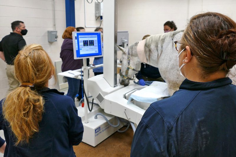
A generous estate gift designated for facility upgrades has enabled the Marion duPont Scott Equine Medical Center to replace its nuclear scintigraphy equipment.
Nuclear scintigraphy, which converts radiation emitted from the patient into images of the skeletal structures, provides a comprehensive look at the areas of injury in the horse. The procedure can check the entire skeleton for abnormalities, including fracture or osteoarthritis, and can pinpoint inflammation due to infection or trauma. The modality is particularly effective at diagnosing difficult lameness issues or sore spots, which are revealed by increased radioactivity.
Although nuclear scintigraphy has been one of the key imaging modalities available at the center since 1995, vast improvements to the technology’s mechanics, electronics, and software algorithms have resulted in faster, clearer image captures.
“Upgraded nuclear scintigraphy equipment broadens our imaging capabilities and adds to an already-extensive suite of imaging modalities, all of which are central to the high level of clinical services that we offer to our clients,” said Michael Erskine, Equine Medical Center director and Jean Ellen Shehan Professor.
To house the new equipment, the center’s designated nuclear scintigraphy room was retrofitted with specialized, non-slip, poured flooring to ensure the safety of horses and staff. On May 7, the newly installed MiE Equine Scanner H.R. was used for the first time.
The scanner’s equine gamma camera is suspended just above the floor on a floating gantry, which allows full mobility of the equipment for optimal positioning, eliminating the need for undue repositioning of the equine patient. Not only can large areas of the horse be imaged at once, real-time motion correction ensures that the images remain sharp, despite any movement by the horse.
During image capture, the equipment’s integrated acquisition and processing system allows live review of the images on two screens, including the ability to compare limb to limb so that the perfect image of an area of interest is obtained.
Able to image literally every inch of a horse, nuclear scintigraphy has proven to be invaluable for diagnosing many orthopedic injuries that are not easily localized using other imaging techniques.
“Our upgrade in nuclear scintigraphy technology will ultimately allow us to image horses faster with improved image quality, expanding the number of scans we can perform daily, as well as fine-tuning our diagnostic capabilities,” said Maureen Kelleher, clinical assistant professor of sports medicine and surgery.
The equine patient is admitted to the center the day before the appointment and is bandaged overnight to maintain a suitable core temperature. The following morning, radioactive isotope is intravenously administered in preparation for the imaging process.
Besides providing top-notch diagnostic imaging, the procedure itself is very safe, radiation levels are extremely low, and horses are caused no distress.
Related Content
-
Article Item