More powerful MRI enhances diagnostic, research capabilities at Veterinary Teaching Hospital
September 5, 2023

The old MRI scanner had reached the end of the line. Rather than merely replace it, the Virginia-Maryland College of Veterinary Medicine decided to make a revolutionary upgrade.
A new Siemens 3-tesla MRI scanner is being installed at the Veterinary Teaching Hospital, set to go online for full diagnostic use this month.
A tesla is a unit of measuring magnetic intensity, and the new scanner has double the power of the Phillips 1.5-tesla model that is being replaced. That will improve image quality and quicken scan time considerably.
“From a day-to-day clinical standpoint, the biggest difference between the 1.5-tesla magnet and a 3-tesla magnet is going to be the speed,” said Richard Shinn, assistant professor of neurology in the Department of Small Animal Clinical Sciences. “It’s about twice as fast, so that means less anesthesia time for the patient and potentially being able to see more patients as well.”
The new scanner is on par with that used in many human medical hospitals and is among the first few installed in a veterinary hospital nationally.
“Our old unit was at the end of its lifespan, and it served us well, but we were losing our ability to keep it up and running,” said Gregory Daniel, professor of radiology in the Department of Small Animal Clinical Sciences. “Getting it fixed was going to be problematic because Phillips would no longer be able to support it, and it had limitations as far as what it was capable of doing. This new unit allows us to expand those capabilities.”
“When you go to replace, you can put in a new 1.5 or you can upgrade to the 3T, which is becoming the new sought-out technology,” said Valerie Vaught, supervisor of diagnostic imaging and support services. ”It definitely leads us to new opportunities and new research capabilities.”
The veterinary hospital chose to go with a Siemens model, rather than another by Phillips, largely to coordinate better with the Fralin Biomedical Research Institute for improved collaboration in research.
“Fralin also has a Siemens magnet, so we can work in conjunction with them with the exact same protocols for research cases,” said Vaught. “You can get images that are similar with different manufacturers, but not exactly the same. This will help us be able to expand our research abilities.”
MRI, or magnetic resonance imagery, is an important tool in diagnostic care of animal patients in the veterinary hospital, complementing radiograph and CT (computerized tomography) technology.
“Unlike radiographs and CT which use X-rays to generate an image, there is no ionizing radiation associated with the MRI unit,” Daniel said. “Its claim to fame is that it has very good soft tissue differentiation, we are able to see various components of soft tissue based upon both their molecular makeup and structural integrity. An X-ray just gives us an image based upon how the tissue absorbs or blocks the radiation that's going through it. When we're looking at soft tissue with MRI we see a variety of different intensities based upon the makeup of the soft tissue, whereas, in our CT or radiograph, that can be all pretty homogeneous and non-differentiated.”
The new MRI will be used heavily in neurology, but also in oncology and orthopedics, among other veterinary specialties.
“I think with our neurology caseload being high and the fact that we are doing leading-edge type research in neurology, it's certainly going to be an important step as the program continues to grow,” Daniel said.
Written by Kevin Myatt, Writer/Editor for the Virginia-Maryland College of Veterinary Medicine.
MRI 8
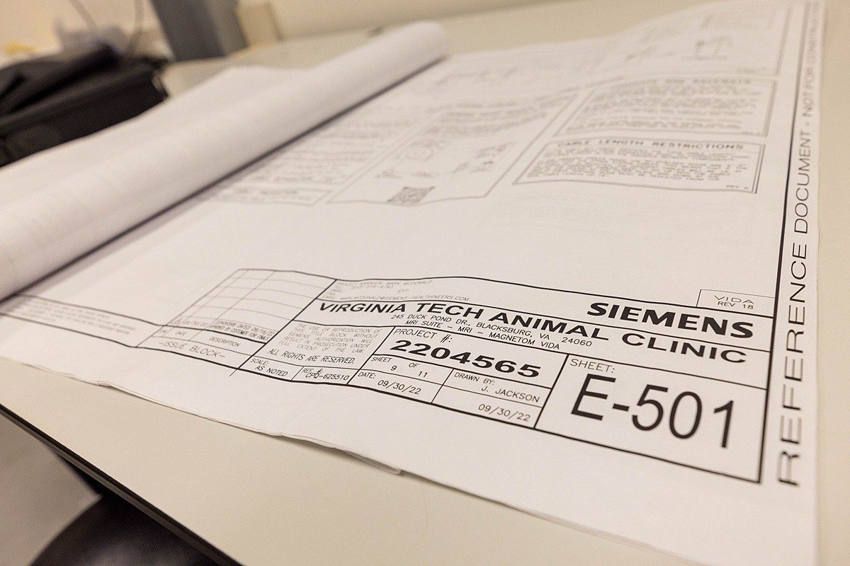
Schematics for the new 3t MRI at the Veterinary Teaching Hospital in Blacksburg, VA. Photo by Andrew Mann for Virginia Tech.
Contact:
Andrew Mann
Director of Communications and Marketing
Related Content
-
Article Item