New table for CT scanner a ‘game-changer’ for examination of large animals at Veterinary Teaching Hospital
July 17, 2023
You can lead a horse to water, but you can’t get one easily into a CT scanner.
Until recently, clinicians at the Veterinary Teaching Hospital (VTH) in the Virginia-Maryland College of Veterinary Medicine couldn’t use CT, or computed tomography, for large animals. But the recent purchase of an $85,000 table for large animals that can be wheeled to the CT scanner now allows for that option, particularly useful for studying conditions inside the heads of equine animals.
“We commonly use the CT scanner for imaging dogs and cats,” said Gregory Daniel, professor of radiology in the Department of Small Animal Clinical Sciences. “The current CT scanner works great for small animals, but most scanners have a 450-pound table limit. For most people and certainly for all small animals, that is sufficient. But if you've got a 1,000-pound patient, then a traditional CT scanner cannot accommodate that weight.”
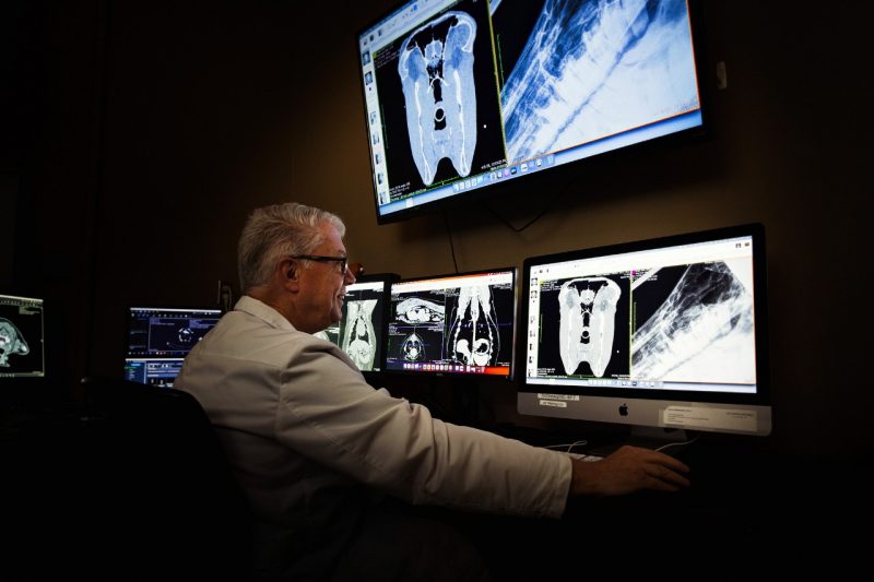
Now, a horse or other large animal can be anesthetized, laid upon the table, and rolled to the CT scanner. The table is electronically synced with the scanner, allowing it to capture slice-by-slice imagery of a large animal’s skull, teeth, and cranial cavities, providing much more detail than X-ray radiographs can reveal.
“When you look at the radiograph of a horse skull, it’s is a two-dimensional image of a three-dimensional object,” Daniel said. “A radiograph is composed of shadows from multiple overlying structures, superimposed over each other. When you’ve got a complex anatomy with a lot of overlying bones and cavities, a radiograph can be challenging to interpret.”
Radiographs work fine for most problems involving teeth or the more outward bone structure, but become more difficult to rely on when issues extend into the nose or other interior parts of a horse’s face and head.
“We do a lot of evaluation of teeth and nasal passages, that's fairly common in horses,” Daniel said. “With traditional radiographs, it's difficult to be precise in the diagnosis. But with CT, we can we can see the structures with far more clarity. And it allows us to be more specific and accurate in determining the underlying cause of a condition.”
VTH in Blacksburg will now be the only veterinary hospital offering the capability to employ CT scans for large animals in a radius extending roughly 240 miles, to as far away as North Carolina State’s veterinary college in Raleigh and to VMCVM’s own Marion duPont Scott Equine Medical Center (EMC) in Leesburg, where there is a CT scanner that allows equine animals to stand during the exam.
“We have referred some of these cases to the EMC because we needed the additional information and that was the best thing for the patient,” said Chris Byron, head of the Department of Large Animal Clinical Sciences. “If they're coming from southern Virginia or North Carolina to us, it's a big ask to send them another three and a half to four hours north. So, for our region, for Southwest Virginia and into West Virginia, it’s certainly a big, big step up in capabilities.”
Byron and Daniel both describe the acquisition of the CT scanner table for large animals as a “game-changer.” Combined with MRI technology that does well with ligaments and cartilage of large animals’ legs and feet, plus existing radiograph and ultrasound abilities, veterinary clinicians at VTH can cover much more of a large animal’s body in examinations.
“Radiographs underestimate the extent of the disease,” Daniel said. “But the CT scanner can greatly improve our ability to diagnose diseases of the head and nasal passages.”